Authors: Tian Shuiqing, Wan Yonghui, Zhou Wei, Shen Yue, Yu Ying, Yang Liting, Xiao Shujun
(Wuhan University People's Hospital, Oncology Center, Wuhan, Hubei 430060, China)
Abstract:
Objective: To explore the application effect of mini-midline catheters in venous therapy for cancer patients.
Methods: From August 2020 to August 2021, 132 cancer patients at a tertiary hospital's oncology center in Wuhan were guided by ultrasound to insert mini-midline catheters, with regular maintenance and observation during catheter retention.
Results: A total of 132 patients were catheterized, with a success rate of 100%. The retention time was 2-44 (16.83±9.12) days; the operation time was 6-40 (14.78±6.14) minutes. The complication rate during catheter retention was 6.8%, with no severe complications such as catheter-related bloodstream infections or thrombosis. After completing treatment, 116 cases were extubated, 7 cases had unplanned extubation due to complications, 1 case had unplanned extubation at night, and 8 cases were extubated due to treatment abandonment or death.
Conclusion: Ultrasound-guided mini-midline catheter insertion has a high success rate, sufficient retention time, low complication rate, and low cost, effectively meeting the needs of some cancer patients for short- to medium-term intravenous infusion therapy, providing a safe venous access throughout their hospitalization.
Keywords: Mini-midline catheter; Cancer; Ultrasound guidance; Venous therapy
Classification Number: R472
Document Identification Code: B
DOI: 10.16460/j.issn1008-9969.2023.04.075
Introduction:
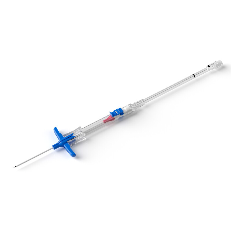
Peripherally inserted central venous catheters (PICC) are the preferred intravenous infusion tool for cancer patients. However, for patients in end-stage palliative care or those requiring short- to medium-term nutritional support, anti-infection, pain relief, and sedation due to severe adverse reactions from chemotherapy and radiotherapy, there is no indication for central venous catheter insertion. Clinically, peripheral venous indwelling needles are often used for intravenous infusion. Studies have shown that compared to PICC and indwelling needles, midline catheters are more suitable for patients hospitalized for 6-14 days, considered the first-line choice for patients with difficult venous access and hospitalization time >48 hours. Peripheral long catheters, also known as mini-midline catheters (Mini-midline catheter or "short" midline catheter), are made of polyurethane, 8-10 cm long, and can be placed in superficial veins of the forearm or arm using direct puncture technique or in deep veins of the upper arm using ultrasound guidance. The tip does not exceed the axilla, meeting the needs of intravenous therapy for ≥1 week. Abroad, they are mainly used in emergency departments, patients with difficult venous access, and palliative care for cancer patients, with good results[5-6]. Domestic research on mini-midline catheters is also gradually gaining attention and importance. Since August 2020, our department has been using mini-midline catheter puncture technology in cancer patients, successfully catheterizing 132 patients with good results, reported as follows.
1. Research Subjects:
Using convenience sampling, 132 cancer patients requiring short- to medium-term intravenous infusion therapy from August 2020 to August 2021 were selected as research subjects. According to the 2021 Infusion Nurses Society (INS) guidelines on the indications for peripheral long catheters, inclusion and exclusion criteria were established. Inclusion criteria: (1) Pathological diagnosis of malignant tumor; (2) Age ≥18 years; (3) Expected infusion cycle of 1-4 weeks; (4) Continuous infusion of isotonic or near-isotonic drugs; (5) Intermittent or short-term infusion of hypertonic, corrosive drugs. Exclusion criteria: (1) Patients undergoing multiple cycles of chemotherapy; (2) Continuous infusion of vesicants, drugs with extreme pH or osmolarity; (3) History of radiotherapy, thrombosis, vascular surgery in the catheterization area, or end-stage renal disease requiring venous protection. This study was approved by the Clinical Research Ethics Committee of Wuhan University People's Hospital (No. WDRY2021-K102), and informed consent was obtained from patients and their families before catheterization.
A total of 132 patients were catheterized, including 79 males and 53 females, aged 24-92 (65.32±12.72) years; 23 had undergraduate education, 51 had junior college education, 35 had secondary school education, 16 had primary school education, and 7 were illiterate. Disease types: 35 cases of lung cancer, 9 cases of intracranial malignant tumors, 28 cases of liver cancer, 6 cases of laryngeal cancer, 3 cases of gastric cancer, 23 cases of nasopharyngeal cancer, 7 cases of rectal cancer, 5 cases of pancreatic cancer, 3 cases of esophageal cancer, 6 cases of cholangiocarcinoma, 2 cases of multiple myeloma, 3 cases of colon cancer, 1 case of prostate cancer, and 1 case of kidney cancer. Among them, 57 patients were in end-stage palliative care, 41 patients were receiving supportive treatment for adverse reactions to chemotherapy and radiotherapy, and 34 patients were undergoing interventional, immunotherapy, or targeted therapy; 81 patients were infused with anti-inflammatory, sedative, analgesic, isotonic or near-isotonic drugs, 12 patients received intermittent short-term infusion of vasoactive drugs, and 39 patients received intermittent infusion of parenteral nutrition, dehydration, and other hypertonic drugs.
2. Methods:
2.1 Operators and Materials:
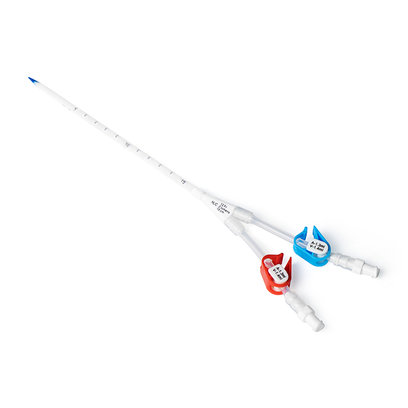
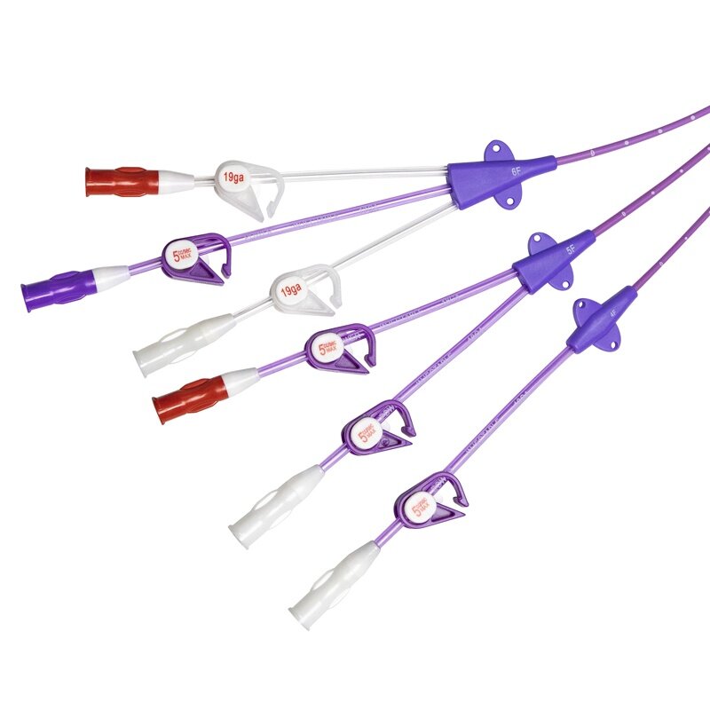
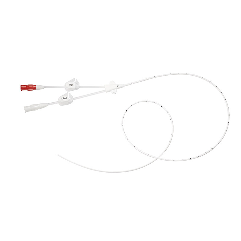
The procedures in this study were performed by two PICC specialist nurses certified by the Chinese Nursing Association, each with over 5 years of experience in ultrasound-guided venous puncture. One specialist nurse had a master's degree, and the other had a bachelor's degree, both were senior nurses with over 10 years of nursing experience and had received training and assessment in vascular ultrasound. The materials used in this study were medium-length catheters produced by Haolang Company, specifications PIV4Fr×10 cm / PIV3Fr×8 cm, model P120-04110; Beijing Zhiying Vascular Puncture Navigation System, model PA12W.
2.2 Catheterization Method:
Ultrasound-guided venipuncture above the elbow was used. (1) Positioning was the same as for PICC puncture. (2) Selection of puncture site and catheter: The puncture point was at least 5 cm above the elbow to avoid affecting limb movement and catheter fixation. At the same time, the position 8 cm/10 cm above the puncture point was located to ensure the catheter tip did not exceed the axilla and to avoid venous valves and branches. According to the principle that the catheter-to-vessel diameter ratio should be ≤45% and the catheter length in the vein should be >2/3 of the catheter length, a 3 F×8 cm or 4 F×10 cm catheter was selected. (3) Establish a sterile area. (4) Prepare items: sterile protective cover for ultrasound probe, active catheter and guide wire, check catheter integrity. (5) Puncture: Use transverse axis puncture, hold the ultrasound probe with the left hand to position the blood vessel image in the center of the ultrasound probe, and hold the catheter handle with the right hand in a pen-like grip. At a position 0.4-0.5 cm from the center of the ultrasound probe, insert the needle at a 45°-60° angle towards the center of the ultrasound probe, aiming for the blood vessel seen under ultrasound. Observe good blood return in the blood return area, reduce the angle and advance the needle about 0.2 cm to ensure the catheter enters the blood vessel. (6) Insert the guide wire and catheter. Compared to the modified Seldinger technique for PICC or ordinary midline catheters, the mini-midline catheter uses a direct Seldinger technique without the need for skin dilation or microvascular sheath replacement. After the puncture needle successfully enters the target blood vessel, the guide wire is directly delivered, and the catheter handle is rotated from the card slot to the guide rail of the handle, slowly pushing the catheter along the guide wire and guide rail to the puncture point. If difficulty or resistance is encountered when inserting the guide wire or catheter, confirm under ultrasound whether the needle tip is in the blood vessel or if it encounters a venous valve or branch, and adjust accordingly without forcing the catheter. (7) Withdraw the needle and guide wire: Press the catheter tip to prevent blood from flowing out of the catheter end, and withdraw the puncture needle and guide wire. (8) Connect the extension tube and infusion connector, flush the catheter with a pulsatile technique, and seal the catheter with positive pressure. (9) Tighten the locking device, remove the catheter handle, and tear off the peelable seat. (10) Fixation: Use a special fixation wing to secure the catheter, apply a sterile dressing, and use tape to cross-fix the tail end in a butterfly shape. (11) Record.
2.3 Maintenance and Quality Control of Mini-Midline Catheters
1. Dressing Change: Unlike PICC or standard midline catheters, mini-midline catheters do not require a dressing change within the first 24 hours post-insertion. Other maintenance frequencies, methods, and standards remain the same.
2. Catheter Fixation: Unlike standard indwelling needles that are fully inserted, mini-midline catheters should be fixed with 0.3-0.5 cm exposed to reduce kinking at the catheter exit site. The U-shaped fixation of the extension tube and positioning the catheter clamp near the catheter hub can reduce blood reflux and prevent occlusion.
3. Flushing and Locking: The method and frequency are the same as for PICC. It is important to use 10 u/mL heparin saline 3-5 mL for positive pressure locking after each flush. For continuous infusion, administer 10-15 mL of 0.9% sodium chloride injection every 6-8 hours using a pulsatile technique. Flush promptly after infusing parenteral nutrition, blood products, and other high-viscosity drugs to reduce the risk of catheter occlusion.
4. Observation Log: Daily observation and documentation of the puncture site, dressing, and local skin condition. Monitor patient complaints for signs of limb swelling, pain, or puncture site exudate.
5. Intermittent Infusion of Vasoactive and Hypertonic Drugs: From the start of infusion, apply Hirudoid ointment along the vein above the puncture site three times a day. Conduct hourly catheter observations and assessments to prevent drug extravasation and phlebitis.
2.4 Observation Indicators
1. Catheterization Operation: Includes the puncture vein, catheter specifications, success rate of single puncture, operation time, and occurrence of arterial puncture or nerve injury. Single puncture success rate: successful entry into the target vein on the first attempt without changing the puncture site. Operation time: the time from vein assessment to catheter fixation completion.
2. Retention Time: The total time from the day of puncture to the day of catheter removal, measured in days.
3. Complications During Retention: Includes local bleeding at the puncture site, exudate, catheter occlusion, phlebitis, catheter-related thrombosis, and puncture site infection. Local bleeding at the puncture site is defined as the presence of blood-tinged fluid exuding from the puncture site. Exudate at the puncture site is defined as the presence of colorless, odorless transparent fluid or pale yellow fluid exuding from the puncture site, with fluid leakage during injection or infusion. Catheter occlusion is defined as a slowed or completely obstructed infusion rate, inability to aspirate blood, or inability to flush the catheter. Phlebitis is defined as inflammation of the vein, clinically manifested as pain/tenderness, erythema, swelling, induration, suppuration, or palpable venous cord formation, evaluated according to the 2021 INS standards into grades 0-IV. Catheter-related venous thrombosis is defined as swelling, redness, and tenderness of the limb on the catheterized side, with Doppler ultrasound showing substantial hypoechoic masses in the vein, significant venous lumen expansion, non-collapse upon probe compression, and absence of significant blood flow signals. Puncture site infection is defined by the presence of redness, swelling, and induration at the catheter entry site, with severe cases showing purulent discharge.
2.5 Statistical Methods
Data processing was performed using EXCEL 2019 software, with descriptive statistics for catheterization conditions, operation time, retention time, and complications.
3. Results
3.1 Catheterization Operation: All 132 patients were successfully catheterized, with a success rate of 100%, and no arterial puncture or nerve injury occurred. The operation time ranged from 6 to 40 (14.78±6.14) minutes. Details of the puncture veins, catheter models, and single puncture success rates are shown in Table 1.
.png)
3.2 Incidence of Complications During Retention
During the retention period, complications occurred in 8 cases (6.1%), including 3 cases of puncture site exudate, 3 cases of phlebitis, and 2 cases of catheter occlusion. No severe complications such as catheter-related bloodstream infections or catheter-related thrombosis were observed.
3.3 Retention Time
The catheter retention time ranged from 2 to 44 days (16.83±9.12 days). Among these, 67 cases had a retention time of 4-15 days, 51 cases had a retention time of 16-30 days, and 14 cases had a retention time of 30-44 days. A total of 116 cases had the catheter removed after completing treatment, 7 cases had unplanned removal due to complications, 1 case had unplanned removal at night, and 8 cases had removal due to patient discontinuation of treatment or death.
4 Discussion
4.1 Ultrasound-Guided Mini-Midline Catheters Improve Success Rate and Shorten Catheterization Time
In this study, all 132 patients were successfully catheterized, with a success rate of 100% and a single puncture success rate of 83.3%, slightly lower than the 97.4% first puncture success rate reported for ultrasound-guided superficial vein indwelling needles. This discrepancy may be due to the novelty of mini-midline catheterization in the country and the catheter's length and vein depth. Mini-midline catheters are 8-10 cm long, with an average vein depth of >0.5 cm, increasing the difficulty of puncture and catheter advancement. However, the single puncture success rate was significantly higher than the 65.52% reported for manual indwelling needle puncture. Ultrasound visualization clearly shows the anatomical structure of the veins, allowing observation of vein direction, branches, and valves, and measurement of the vein lumen diameter and depth from the body surface, reducing catheterization failure due to large catheter-to-vein ratio. Ultrasound guidance enables precise puncture of the target vein, tracking the needle tip and catheter, and confirming the catheter's position within the vein, thus improving puncture success rates. The 2021 INS guidelines recommend using ultrasound for placing long peripheral catheters in adults and children with difficult peripheral veins to improve first puncture success rates and reduce operation time. The operation time for mini-midline catheters was 6-40 minutes (14.78±6.14 minutes), similar to the 14±6 minutes reported by Magnani et al. for ultrasound-guided mini-midline catheterization in palliative care patients, and significantly shorter than the average 35 minutes for modified Seldinger technique. The integrated structure of the mini-midline catheter's needle, guidewire, and catheter, combined with ultrasound visualization, simplifies and speeds up the procedure, eliminating the need for X-ray positioning and significantly shortening catheterization time compared to traditional midline catheter modified Seldinger technique. This provides rapid venous access for critically ill patients and those with difficult venous access, saving manpower, material, and financial resources.
4.2 Mini-Midline Catheters Reduce Peripheral Vein-Related Complications
The complication rate during catheter retention was 6.8%, with no severe complications such as catheter-related bloodstream infections or thrombosis. This is consistent with international reports on mini-midline catheter use, primarily showing phlebitis, catheter occlusion, and extravasation/exudation, and lower than the 8.7% complication rate reported for ultrasound-guided traditional midline catheters in AIDS patients, and much lower than the 56.14% complication rate for manual midline catheter puncture. The incidence of mini-midline catheter complications is related to vein selection, drug properties, and catheter length. Studies show that 85% of peripheral vein complications occur in peripheral infusion catheters in the distal antecubital fossa of the upper limb, where veins are small and fragile, prone to rupture and extravasation. An ICU study found a 50% risk of infiltration and extravasation within 24 hours for upper arm vein indwelling needles guided by ultrasound, likely due to insufficient catheter length (<5.2 cm). Unlike standard peripheral indwelling needles, mini-midline catheters are longer (8-10 cm), providing better stability for obese, edematous, and deep-vein patients, reducing the risk of drug extravasation and catheter dislodgement. Mini-midline catheters also have a wider lumen, terminating in larger peripheral veins, with the catheter tip located in the axillary vein of the upper arm, where blood flow is faster (300 mL/min), ensuring adequate drug dilution and significantly lower rates of phlebitis and thrombosis.
In this study, all 3 cases of phlebitis were associated with cephalic vein puncture. Previous studies indicate that the incidence of complications is lower with basilic vein puncture compared to cephalic and brachial veins when using mini-midline catheters, suggesting basilic vein should be the first choice. Puncture site exudate occurred in 3 cases on the 7th and 10th days of catheter retention, respectively. These patients received parenteral nutrition and hypertonic drugs, and were third-puncture success cases. Repeated punctures damage the vascular endothelium, and high-concentration parenteral nutrition increases blood viscosity and plasma osmolality, leading to dehydration of vascular endothelial cells, blood cell aggregation near the catheter tip, vein narrowing, or thrombosis, restricting blood flow and causing infusion solution leakage from the puncture site. Therefore, strict adherence to indications is necessary, prohibiting continuous infusion of hypertonic and highly irritating drugs through mini-midline catheters to prevent drug extravasation and thrombosis. For intermittent infusions, preventive measures such as warm compresses and Hirudoid ointment application are recommended. Two cases of catheter occlusion were reported, one resolved with urokinase, and one required catheter removal due to ineffective thrombolysis. The open-tip design of mini-midline catheters, high blood flow in the deep veins of the upper arm, blood pressure measurement on the catheterized limb, vigorous exercise, and heavy lifting can all promote blood reflux and catheter occlusion. Nurses should provide thorough patient education, closely monitor and carefully hand over shifts, promptly and correctly flush and lock the catheter upon detecting blood reflux, and properly close the catheter clamp to prevent occlusion.
4.3 Mini-Midline Catheters Provide Adequate Retention Time for 1-4 Weeks of Intravenous Therapy
The retention time for mini-midline catheters in this study was 2-44 days (16.83±9.12 days). Patients receiving palliative sedation and analgesia for advanced cancer, interventional therapy, immunotherapy, targeted therapy, and those requiring nutritional support due to oral ulcers and swallowing difficulties in the late stages of head and neck radiotherapy typically require 1-4 weeks of treatment. Peripheral vein indwelling needles are commonly used due to cost and patient preference, despite high rates of phlebitis and extravasation, causing significant patient discomfort from frequent replacements. While PICC and infusion ports are options, they are associated with short- and long-term complications, complex procedures, and high costs. The 2021 INS guidelines recommend mid- to long-term catheters or long peripheral catheters for patients with expected treatment cycles of 1-4 weeks and peripheral veins that can tolerate the drug properties. Mini-midline catheters, due to their length, placement in deep veins of the upper arm, and tip location in fast-flowing peripheral veins, have a low complication rate, ensuring longer retention times and meeting short- to mid-term intravenous therapy needs. Although traditional midline catheters are also suitable for treatments exceeding one week, the direct Seldinger technique for mini-midline catheters makes puncture more convenient and faster, reducing contamination and infection risks. Additionally, mini-midline catheters are more cost-effective than PICC, costing only one-third of PICC prices, reducing patient medical expenses while ensuring safety and reliability, making them a viable short- to mid-term intravenous therapy alternative.
5 Conclusion
The application of mini-midline catheters expands the range of intravenous infusion tools for cancer patients, offering high catheterization success rates, short operation times, and low costs, with a low incidence of complications during retention. They meet the infusion needs of patients requiring ≥1 week of treatment and can be a preferred intravenous therapy tool for palliative care in terminal cancer patients or routine intravenous therapy and intermittent short-term infusion of irritating drugs without central venous catheter indications. The limitations of this study include the sample not fully representing the entire cancer patient.

.png)